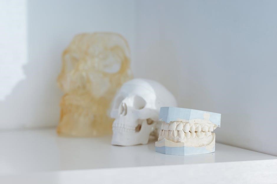
The skull‚ part of the axial skeleton‚ comprises 22 bones forming the cranium and face. It protects the brain‚ houses sensory organs‚ and showcases anatomical complexity‚ vital in medical studies.
Overview of the Skull Structure
The skull is a complex skeletal structure composed of 22 bones that form the cranium and facial skeleton. It is divided into two main parts: the cranial skeleton‚ which encloses and protects the brain‚ and the facial skeleton‚ which supports facial features and functions. The cranial skeleton consists of 8 bones‚ including the frontal‚ parietal‚ occipital‚ temporal‚ sphenoid‚ and ethmoid bones‚ forming a protective vault around the brain. The facial skeleton‚ made up of 14 bones‚ includes the maxilla‚ zygomatic‚ nasal‚ lacrimal‚ and mandible bones‚ which facilitate functions like chewing‚ breathing‚ and facial expressions. The skull base‚ particularly the occipital bone‚ forms the posterior cranial fossa‚ while the sphenoid bone contributes to the anterior cranial fossa. The skull also features numerous foramina and fissures that allow nerves and blood vessels to pass through‚ ensuring vital connections between the brain and other body systems. This intricate structure highlights the skull’s dual role in protection and functional support.
Importance of Skull Anatomy in Medical Studies
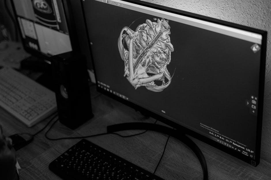
Understanding skull anatomy is fundamental in medical studies due to its critical role in neurology‚ surgery‚ and radiology. The skull’s complex structure‚ comprising 22 bones‚ protects the brain and houses sensory organs‚ making it essential for diagnosing and treating head injuries. Knowledge of cranial anatomy aids in identifying fractures‚ abnormalities‚ and surgical access points‚ such as the anterior and posterior cranial fossae. Foramina and fissures in the skull‚ like the foramen magnum‚ are vital for nerve passage and surgical planning. Skull anatomy also informs neurosurgical procedures‚ such as craniotomies‚ and helps in understanding cranial development anomalies. Additionally‚ it is crucial for interpreting radiological images‚ enabling accurate diagnoses of conditions like fractures or tumors. This knowledge is indispensable for medical professionals‚ ensuring precise and effective patient care in neurosurgery‚ ENT‚ and trauma cases.
Bones of the Cranium
The cranium consists of eight bones—frontal‚ parietal‚ occipital‚ temporal‚ sphenoid‚ and ethmoid—forming the cranial skeleton. These bones provide structural support and protection‚ each contributing unique features to the cranial cavity.
Frontal Bone
The frontal bone forms the upper part of the cranium‚ creating the forehead and the upper portion of the eye sockets. It is a single‚ flat bone with a squamous part and two orbital portions. The frontal bone also contributes to the anterior cranial fossa‚ housing the frontal lobe of the brain. Key features include the supraorbital foramen‚ which allows nerves and vessels to pass through‚ and the frontal sinuses‚ air-filled cavities that reduce the skull’s weight. This bone articulates with the parietal bones at the coronal suture and with the nasal and lacrimal bones in the facial region. Its robust structure provides protection for the brain while supporting facial structures. Understanding the frontal bone’s anatomy is crucial in neurosurgery and craniofacial reconstruction‚ as it plays a vital role in both cranial and facial integrity.
Parietal Bones
The parietal bones are a pair of flat‚ curved bones located on the top and sides of the cranium. They form the roof and upper lateral walls of the cranial cavity‚ providing protection for the brain. Each parietal bone has a squamous part and a tubular part‚ with the squamous portion articulating with the frontal bone at the coronal suture. The parietal bones also meet at the sagittal suture‚ forming the midline of the skull. Key features include the parietal foramen‚ which transmits nerves and blood vessels‚ and the temporal lines‚ where muscles of mastication attach. The parietal bones contribute to the cranial vault’s strength and stability while maintaining the brain’s protective environment. Their anatomy is significant in neurosurgery and cranial reconstruction‚ as they are often involved in surgical access to intracranial structures. Understanding their structure and articulations is essential for medical professionals studying skull anatomy.
Occipital Bone
The occipital bone is located at the posterior aspect of the skull‚ forming the back and base of the cranium. It is a single bone that creates the posterior cranial fossa‚ which houses the cerebellum and the brainstem. A key feature of the occipital bone is the foramen magnum‚ a large opening through which the spinal cord connects with the brain. The bone also includes the external occipital crest and the external occipital protuberance‚ which serve as attachment points for muscles‚ such as the occipitalis muscle‚ facilitating head movement. The occipital bone articulates with the parietal bones along the lambdoid suture‚ contributing to the structural integrity of the skull. Its internal surface is smooth‚ while the external surface is more rugged‚ providing anchors for muscles and ligaments. The occipital bone plays a vital role in protecting the brain and supporting the skull’s posterior structure.

Temporal Bones
The temporal bones are a pair of complex bones located on the lateral aspects of the skull‚ contributing to both the cranial and facial structures. Each temporal bone consists of two main parts: the squamous part and the petrous part. The squamous part forms the lateral wall of the cranium‚ while the petrous part is denser and houses the inner ear structures‚ including the cochlea and semicircular canals. Key features of the temporal bone include the mastoid process‚ a bony projection behind the ear‚ and the styloid process‚ which serves as an anchor point for muscles and ligaments. The temporal bone also contains the external auditory meatus‚ leading to the eardrum‚ and forms part of the zygomatic arch‚ which supports the cheekbones. The temporal bones articulate with the parietal‚ occipital‚ and sphenoid bones‚ playing a crucial role in the structural integrity of the skull while protecting the sensory organs of hearing and balance.
Sphenoid Bone
The sphenoid bone‚ situated in the floor of the cranial cavity‚ is a butterfly-shaped bone that forms a critical connection between the cranial and facial structures. It is located posterior to the frontal bone and anterior to the occipital bone‚ lateral to the ethmoid bone‚ and medial to the temporal bones. The sphenoid bone consists of a central body‚ two greater wings‚ and two lesser wings. The body of the sphenoid contains the sella turcica‚ which houses the pituitary gland‚ and forms part of the anterior cranial fossa. The greater wings project laterally‚ contributing to the lateral walls of the cranial cavity and forming part of the orbit. The lesser wings form part of the anterior cranial fossa and the orbit. The sphenoid bone also contains several important foramina‚ including the foramen ovale‚ foramen spinosum‚ and foramen rotundum‚ which transmit nerves and blood vessels essential for facial and cranial functions. Its complex structure supports cranial stability while facilitating neural and vascular connections.
Ethmoid Bone
The ethmoid bone is a small‚ irregular‚ spongy bone located at the roof of the nasal cavity and the floor of the anterior cranial fossa. It forms part of the anterior cranial base and is positioned between the nasal cavity and the orbit. The bone is divided into three main parts: the perpendicular plate‚ the orbital plate‚ and the lateral mass. The perpendicular plate forms the superior portion of the nasal septum‚ while the orbital plate contributes to the medial wall of the orbit. The lateral mass contains numerous small air cells‚ known as ethmoidal air cells‚ which lighten the bone and contribute to the paranasal sinus system. The ethmoid bone also features important structures such as the superior and middle nasal conchae‚ which project into the nasal cavity and support the mucous membranes. Its complex structure includes foramina for nerves and blood vessels‚ such as the ethmoidal canals‚ and articulates with the frontal‚ sphenoid‚ and nasal bones to form a vital part of the cranial and facial anatomy.
Facial Bones and Their Functions
The facial bones include the maxilla‚ zygomatic‚ nasal‚ and lacrimal bones‚ forming the upper jaw‚ palate‚ and orbital structure. They support facial features‚ enable chewing‚ and house sensory organs.
Maxilla Bones
The maxilla bones are a pair of large‚ complex bones that form the upper jaw‚ palate‚ and orbital floor. They are crucial for facial structure‚ anchoring the upper teeth and supporting the nasal cavity. The maxilla bones fuse during growth‚ forming a single structure in adulthood. They contain the maxillary sinuses‚ the largest paranasal sinuses‚ which play a role in voice resonance and mucus drainage. The alveolar process of the maxilla holds the upper teeth‚ while the palatine process forms the anterior part of the hard palate. These bones also contribute to the formation of the orbit‚ working in conjunction with other facial bones. Their intricate design allows for both structural support and functional versatility‚ making them essential in craniofacial anatomy and surgery.
Zygomatic Bones
The zygomatic bones‚ commonly known as the cheekbones‚ are paired bones that form the prominence of the cheeks and contribute to the lateral walls and floor of the orbit. They are crucial for facial structure‚ connecting with the frontal‚ maxilla‚ temporal‚ and sphenoid bones. The zygomatic bones also form part of the zygomatic arch‚ which is important for muscle attachments‚ particularly the masseter muscle involved in mastication. Their orbital surface helps form the floor and lateral wall of the eye socket‚ providing structural support for the orbit. These bones also contain the zygomaticofacial and zygomaticotemporal canals‚ which transmit nerves and blood vessels; Their role in both protecting the eye and contributing to facial aesthetics makes them significant in craniofacial anatomy and reconstructive surgery.
Nasal Bones
The nasal bones are a pair of small‚ rectangular bones located at the top of the nose‚ forming the bridge of the nasal structure. They are part of the facial skeleton and contribute to the formation of the anterior part of the nasal cavity. These bones articulate with the frontal bone superiorly and the maxilla bones laterally and inferiorly. The nasal bones are thin‚ flat‚ and slightly curved‚ providing structural support to the nose while forming the anterior nasal aperture. They also serve as attachment points for nasal cartilages and muscles‚ aiding in the shape and function of the nose. Injuries to the nasal bones are common due to their exposed position‚ often resulting in fractures. Understanding their anatomy is crucial for rhinoplasty and reconstructive surgeries. The nasal bones play a vital role in both the aesthetic and functional aspects of the face‚ supporting respiratory and olfactory functions. Their precise alignment ensures proper nasal airflow and symmetry.
Lacrimal Bones
The lacrimal bones are small‚ thin bones located in the medial wall of the orbit. They are paired bones‚ with one located in each orbital cavity. The lacrimal bone forms part of the anterior portion of the orbit and plays a crucial role in the tear drainage system. It contains the lacrimal groove‚ which‚ when combined with the lacrimal groove of the maxilla‚ forms the nasolacrimal canal. This canal allows tears to drain from the eye into the nasal cavity. The lacrimal bone articulates with the frontal bone superiorly‚ the ethmoid bone posteriorly‚ and the maxilla bone inferiorly. Its thin structure makes it one of the most delicate bones in the skull. The lacrimal bone’s position and function are vital for maintaining proper tear drainage and eye health. Understanding its anatomy is essential for ophthalmological and maxillofacial surgeries. Its precise articulation with surrounding bones ensures the structural integrity of the orbital region.